
Zoology - Digestion and Absorption Question with Solution | TestHub
Observe the given diagram and choose the correct option: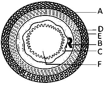
Options:
Answer:
Solution:
The wall of alimentary canal from oesophagus to rectum possesses four layers namely serosa, muscularis, sub-mucosa and mucosa. Serosa is the outermost layer and is made up of a thin mesothelium (epithelium of visceral organs) with some connective tissues. Muscularis is formed by smooth muscles usually arranged into an inner circular and an outer longitudinal layer. An oblique muscle layer may be present in some regions. The submucosal layer is formed of loose connective tissues containing nerves, blood and lymph vessels. In duodenum, glands are also present in sub-mucosa. The innermost layer lining the lumen of the alimentary canal is the mucosa. This layer forms irregular folds (rugae) in the stomach and small finger-like foldings called villi in the small intestine. The cells lining the villi produce numerous microscopic projections called microvilli giving a brush border appearance. These modifications increase the surface area enormously. Villi are supplied with a network of capillaries and a large lymph vessel called the lacteal. Mucosal epithelium has goblet cells which secrete mucus that help in lubrication. Mucosa also forms glands in the stomach (gastric glands) and crypts in between the bases of villi in the intestine (crypts of Lieberkuhn). All the four layers show modifications in different parts of the alimentary canal.
The partis mucosa and in duodenum Brunner's glands are present in submucosa. Parthas longitudinal muscles while Parthas circular muscles which help in peristaltic movements of the alimentary canal. Partis made up of loose connective tissue containing nerves, blood and lymph vessels.